

Research
1. Searching for new early detection markers of OED and OSCC using liquid-based cytology methods
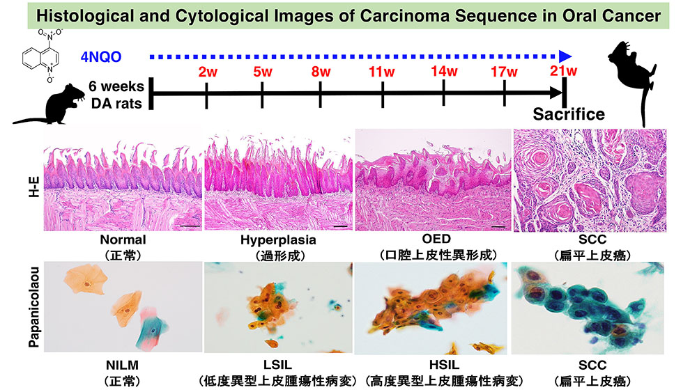
Most oral squamous cell carcinomas (OSCCs) arise from a premalignant lesion, oral epithelial dysplasia; however, useful markers for the early detection of OSCC are lacking. Our study aimed to establish a novel experimental model to observe changes in the sequential expression patterns of mRNAs and proteins in a rat model of tongue cancer using liquid‑based cytology techniques. This novel experimental model allowed the observation of sequential morphological changes and the expression patterns of mRNAs and proteins during carcinogenesis. Combining immunocytochemistry with cytology‑based diagnoses may potentially improve the diagnostic accuracy of OSCC.
2. Demonstration of anti-inflammatory action of Kampo medicines for oral cancer-related cachexia (Improvement of Sarcopenia)
Our study aims to improve cachexia and sarcopenia by using an animal model of tongue cancer and administering herbal medicines to oral cancer-related cachexia. Using our animal model of oral carcinogenesis, we will administer several herbal medicines that improve nutritional, inflammatory, and immune statuses from the time when the development of oral carcinogenesis is confirmed by the LBC method, and confirm the improvement status of anti-inflammatory effects by blood sampling and the LBC method to find the most promising herbal medicines.
3. Survival of salivary pleomorphic adenoma cells in hypoxic condition: analysis by SM-AP cell systems
Among head and neck tumors, pleomorphic adenoma is a benign salivary gland tumor, clinically it is known to recur after surgery or metastasize and develop into a secondary cancer called focal carcinoma. We previously demonstrated that the stromal architecture of pleomorphic adenomas is characterized by hypovascularity, hypoxia, and abundant accumulation of extracellular matrix (ECM) molecules. However, the significance of ECM-rich stroma in the function of salivary pleomorphic adenoma cells remains unknown. In this study, we investigated the hypothesis that the stroma of salivary gland pleomorphic adenomas is hypoxic, enabling tumor cell proliferation without blood supply due to hypovascularization and ECM enrichment in SM-AP cell systems, which are cloned from pleomorphic adenoma of the parotid gland.
4. Histological and proteomic analysis of progression front of oral squamous cell carcinoma
Histopathologically, oral squamous cell carcinoma forms a distinct interface with the surrounding non-cancerous mucosal epithelium. By performing morphological and comprehensive protein analyses of the interface, we identified proteins specifically increasing at the interface between the cancer tissue and the non-cancerous mucosal epithelium. to visualize the differences between cancer cells and non-cancerous cells histopathologically and to elucidate the mechanisms of cancer cell progression. Among the proteins identified, we found that ladinin-1 could related to the directionality of cancer cell progression, and analysis is currently ongoing.
5. Alternative splicing of mRNA in head and neck squamous cell carcinoma
Cancer cells express more diverse proteins than non-cancer cells. One of the mechanisms that generate protein diversity is alternative splicing of mRNA (messenger RNA). Alternative splicing is known to be involved in cancer development/progression and potentially patient prognosis. We are aiming to elucidate the significance of alternative splicing in cancer by integrated analysis using public database search and RNA sequencing using long-read next-generation sequencers. (In collaboration with Prof. Okuda and Assistant Prof. Ling, Niigata University Medical AI Center; Prof. Koelzer and Dr. Lafarge, University Hospital Zurich).
6. Cell death-induced mechanisms for cancer progression
While cancer cells proliferate faster than normal cells, they die more frequently. Chemotherapy and radiotherapy also cause the death of cancer cells. Dead cancer cells are usually cleared by immune cells like macrophages. However, the effect of dead cancer cells on surrounding live cancer cells is not yet fully understood. A previous study revealed that live cancer cells ingest dead cancer cells, which increases the movement of cancer cells and contributes to the progress of cancer. Currently, we are investigating this mechanism in greater detail to develop new therapies that target the role of dead cells in the progression of cancer.